Judging the quality of fused alumina hinges on a comprehensive evaluation of its chemical purity, physical properties, microstructure, and process consistency. High-quality fused alumina is not only pure in composition but also dense in structure and uniform in particle size, significantly improving the performance and lifespan of end products.
Chemical Composition: Purity and Impurity Control are Primary Indicators
Higher Al₂O₃ content indicates better quality:
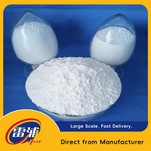
White fused alumina: Al₂O₃ ≥99%, extremely low impurities, suitable for precision machining and high-end ceramics.
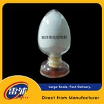
Sub-white fused alumina: Al₂O₃ ≥98.5%, between white and brown fused alumina, offering high cost-effectiveness.
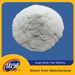
Brown fused alumina: Al₂O₃ approximately 76%~90%, contains more impurities such as Fe₂O₃ and SiO₂, suitable for general industrial applications.
Key impurities must be strictly controlled:
Na₂O: ≤0.3%~0.5%. Excessive levels can cause the formation of the β-Al₂O₃ phase at high temperatures, affecting stability and thermal shock resistance.
Fe₂O₃, SiO₂, CaO: These impurities lower the melting point, increase the glassy phase, and affect hardness and corrosion resistance.
These can be detected using methods such as ICP-OES/MS and XRF. For high-purity products, it is recommended to use ICP-MS with a detection limit at the ppb level.
Microstructure: Reflects the intrinsic quality of the material
Crystal morphology and size: High-quality fused alumina should be polygonal granular, with large crystals (100–250 μm) and tightly packed, achieving the densest possible packing.
The presence of numerous small pores or loose, foamy structures (such as edge material) indicates inferior quality.
Crystal Phase Composition: The main crystal phase is α-Al₂O₃ (corundum phase), the content of which can be confirmed by XRD analysis.
Transitional phases such as γ-Al₂O₃ should be avoided due to their poor thermal stability.
Microscopic Defects: SEM (Scanning Electron Microscopy) should be used to observe for intergranular cracks, inclusions, or unmelted particles ("under-melted material").
High-quality materials should be free of obvious layered or lamellar aggregates; a step height of 0.2–1.0 μm is within the normal range for step growth morphology.